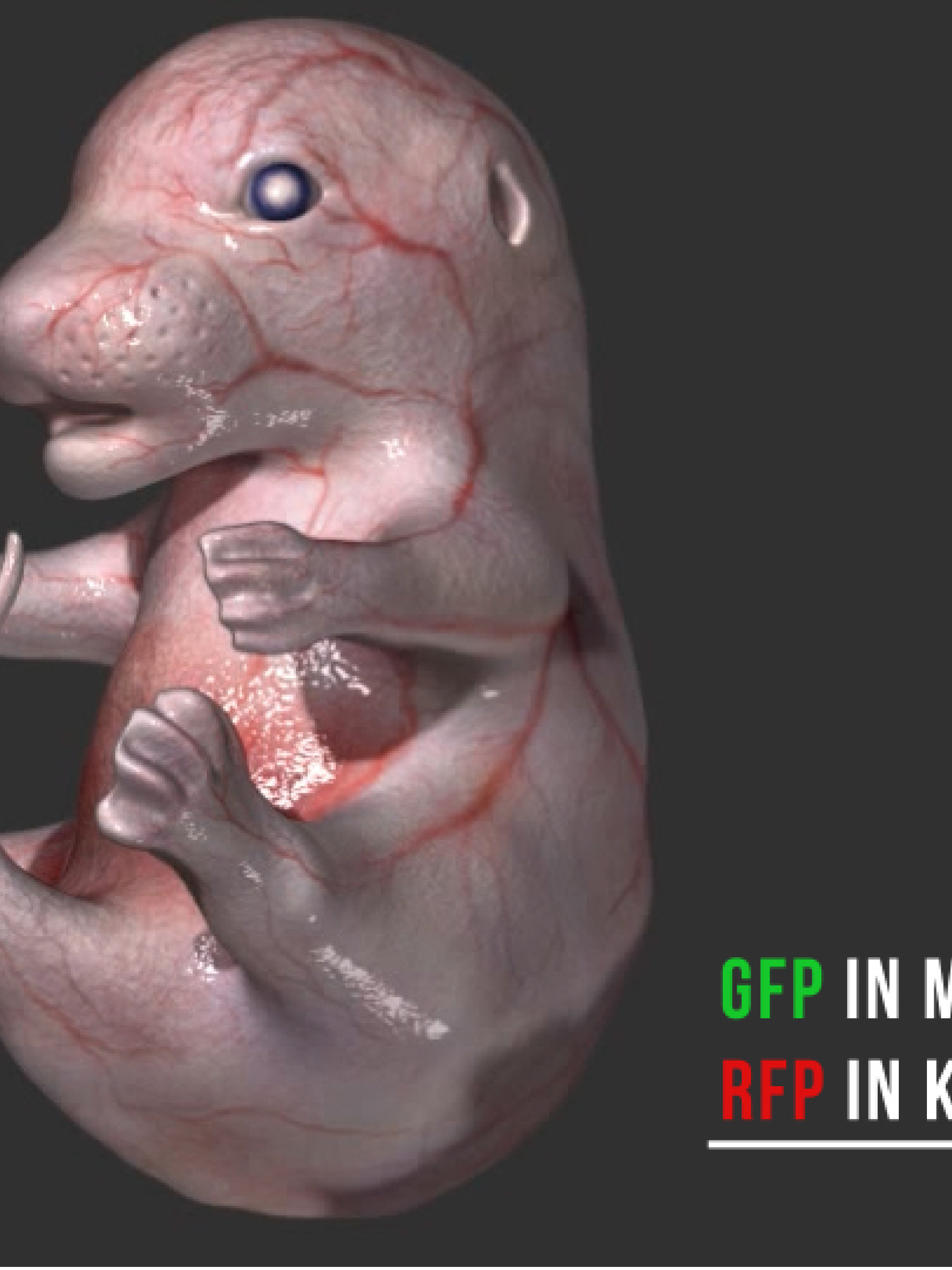
This 3D animation describes the imaging of skin cells using confocal microscopy. Research into how cells move is important for understanding skin cancer and to discover new ways to fight it.
Zbrush and Maya were used to create the 3D models, and Adobe After Effects to compile the final animation.
This is an excerpt from a work in progress collaboration with a scientist at the CRUK Beatson Institute studying how cells respond to chemical signals in their environment.
Created in Maya and Adobe After Effects.
A simple animation explaining different types of blood cells.
Created in Maya and Adobe After Effects.
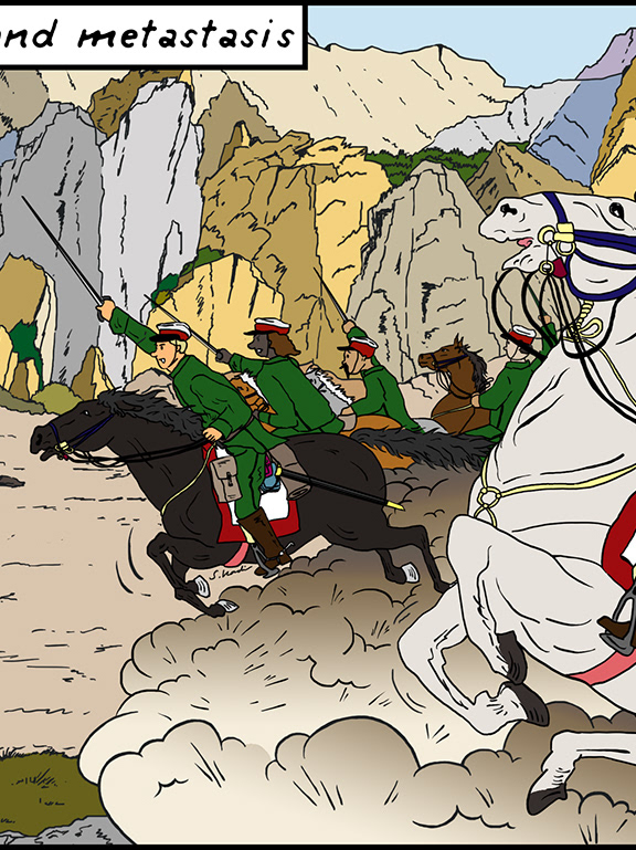
This was the animation I made for my MSc in Medical Visualisation and Human Anatomy at the Glasgow School of Art and Glasgow University. It was a collaboration with Professor Insall at the CRUK Beatson Institute to explain two different hypotheses of cell migration and the role of a well studied cell protein called ‘actin’.
Software used: Zbrush, Maya, Adobe After Effects and Audition.
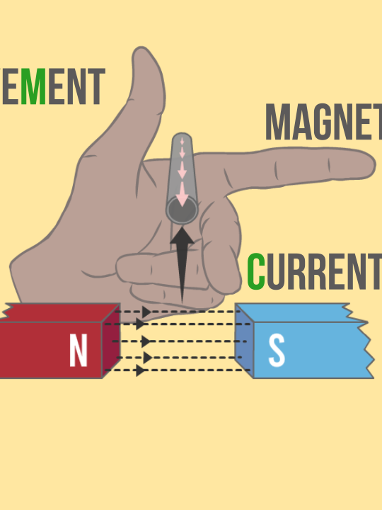
A short introduction about membrane receptors in the style of cut out animation.
Created using Adobe Illustrator and After Effects.
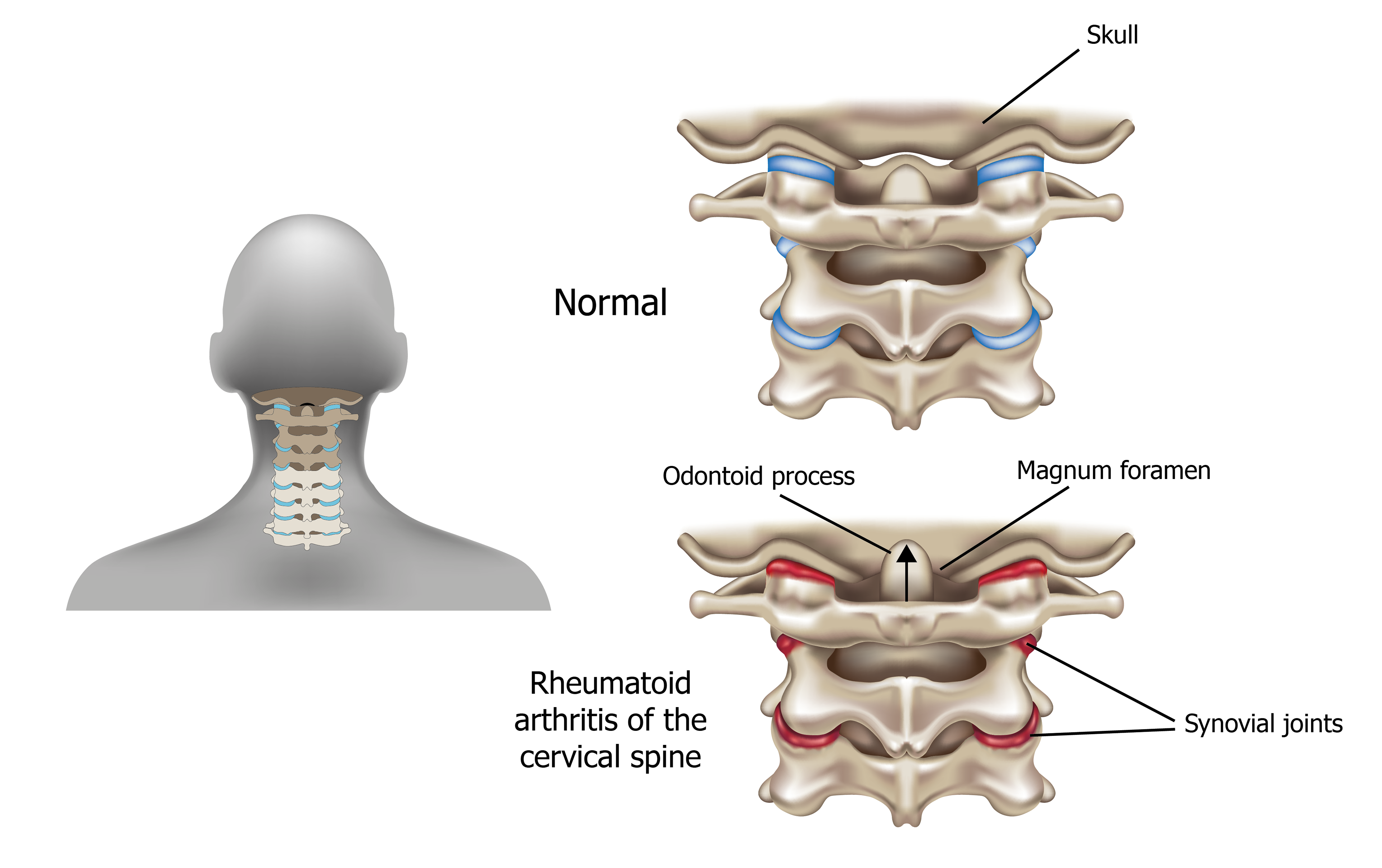
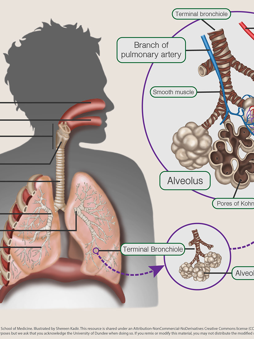
During my graphic design internship at Costello Medical I was given the opportunity to make scientific illustrations depicting
the effects of rheumatoid arthritis. I used Adobe Illustrator to create the image above to highlight the damage the disease
does to the bones and joints in the cervical spine.
the effects of rheumatoid arthritis. I used Adobe Illustrator to create the image above to highlight the damage the disease
does to the bones and joints in the cervical spine.
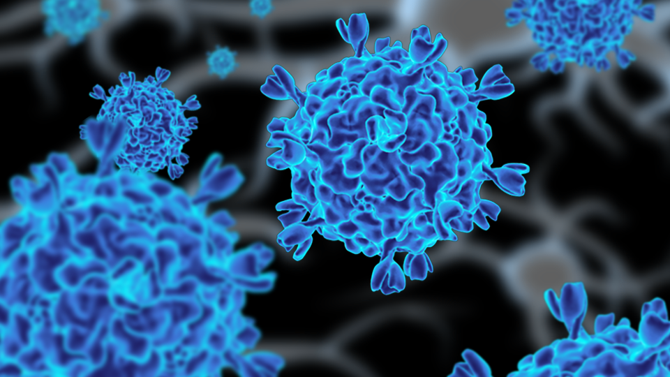
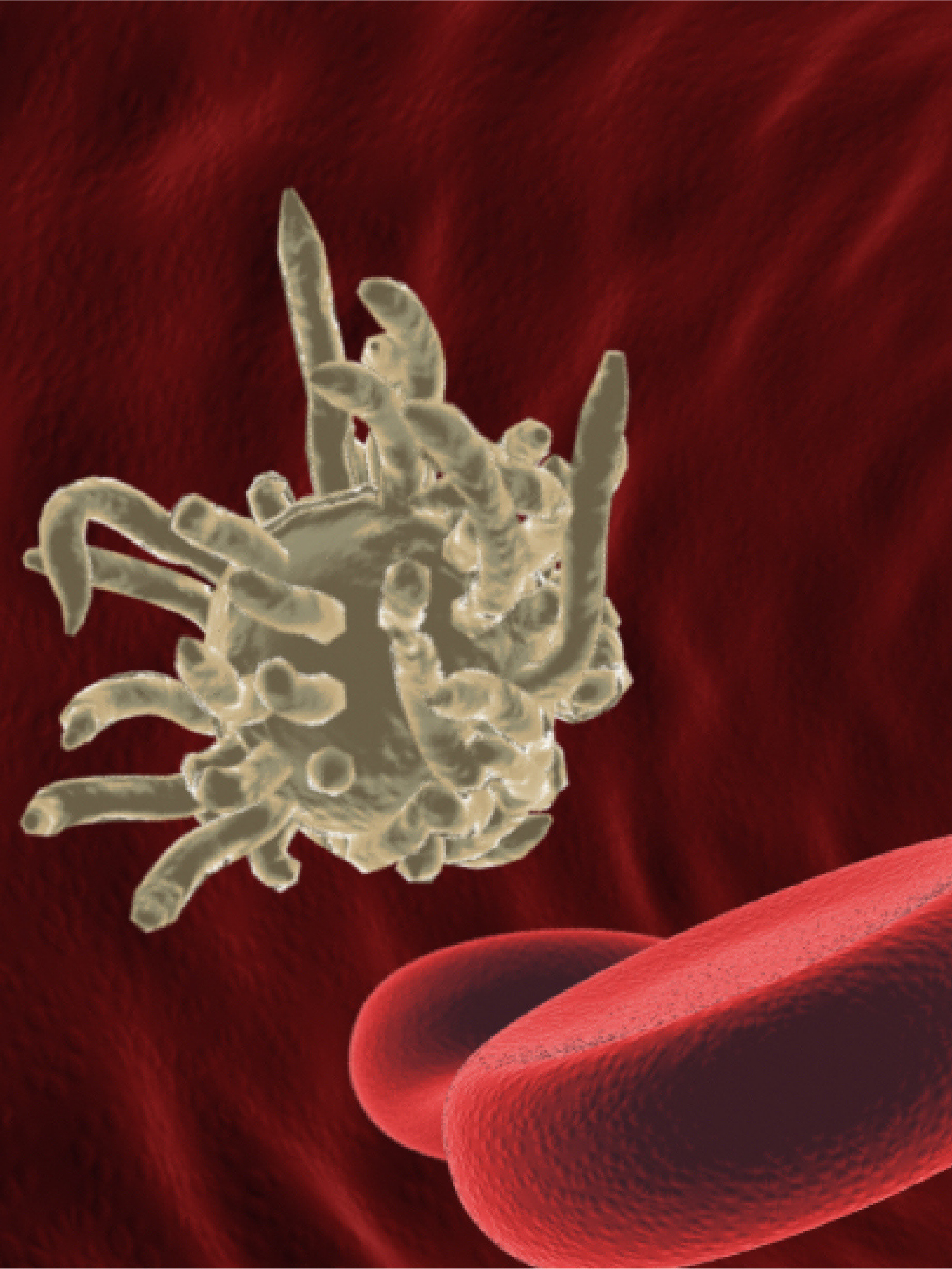
Viral infection illustration.
I used the plugin mMaya to download the structure of the spiroplasma virus from the protein data bank. I textured and rendered the virus in Maya and the final composition was done in Adobe Photoshop.